Clinical Cases:
Endodontic
treatments

Endodontics is the branch of dentistry that has as its objective of study the tissues located inside of the tooth (dental pulp or nerve) and the treatment of the diseases related to them.

Endodontic treatment is commonly associated, although inappropriately, with the term “tooth killing”. It is a dental surgery that has as its main purpose the protection of the tooth and it tries to avoid as much as possible turning to extraction and implantology.

Performing correct endodontic treatment, using cutting-edge materials and toolkits (an operating microscope, tools made of special alloys, innovative biomaterials, etc.), is of paramount importance for the teeth to continue to carry out the normal masticatory function without presenting in the future any complications such as acute abscesses or granulomas.

The patient must, therefore, be aware that endodontic treatment is far from being an ordinary medical solution that is easy to perform, as this is often perceived from the result of a widespread lack of understanding and a very vague approach expressed by some operators. This can even result in the patient having misconceptions about the costs of endodontic treatment, given that often it is believed to be inexpensive, especially compared to implants that have, sometimes unjustifiably, very high prices.

In recent years there has in fact been a strong and objectionable increase in dental replacements with implants, a clear signal that the approach to quality endodontics is unfortunately still not widespread.

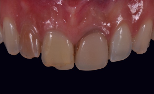
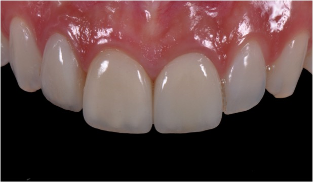
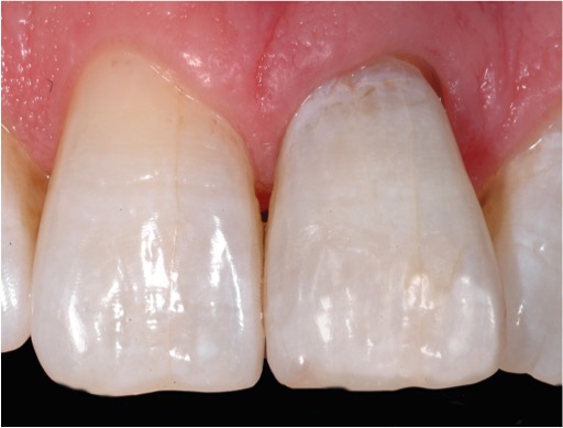
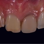
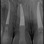
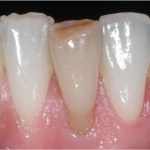
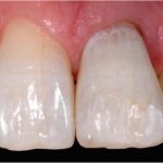
Chromatic alteration of the dental elements affected by endodontic problems.

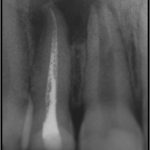
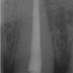
In detail we note an apical radiolucency caused by the necrosis of the central incisor and by the incongruous root canal treatment of the lateral incisor.

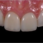
Later, the resolution of the endodontic pathology thanks to a perfect sealing of the root canals, and consequently the resolution of the aesthetic problem with the application of ceramic crowns.

Discover the details

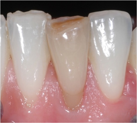
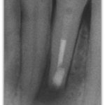
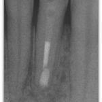
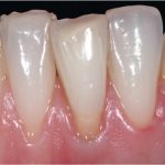
Pulsing necrosis of the lower left central incisor with chromatic alteration.

In detail we note the radiological evidence of bone lesion resulting from necrosis.

At first there was an attempt at grouting therapy which did not have a good end result.
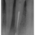
Subsequently, we note the post operative radiograph after apicectomy and the control radiograph after 4 years where the complete recovery of the periapical pathology is visible.

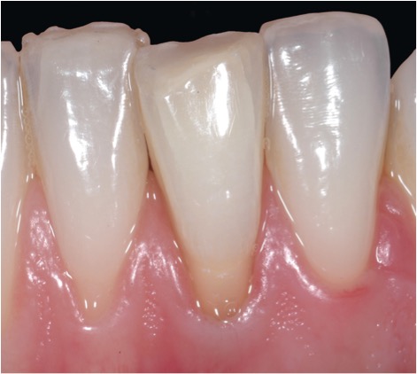
The last photo shows the moment after surgical endodontic therapy and endodontic excavation.

Discover the details

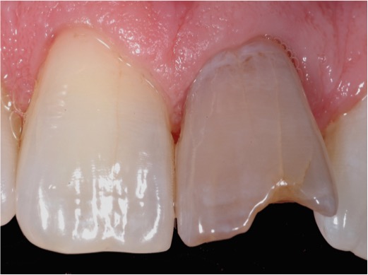
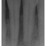
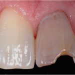
Necrosis of the upper left incisor with discoloration and the presence of large bone rarefaction.

We can see in detail  that there is a dark halo around the root: the sign of a large bone loss around it.

Subsequently there is healing of periapical pathology with complete reformation of the periapical bone.

In the photo at the end of the treatment, the endodontic fading and the restoration with composite resin are visible.

Discover the details

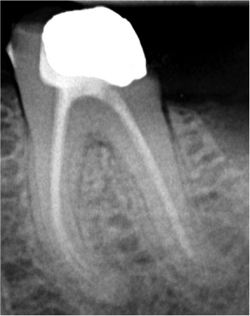
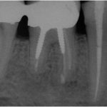
Granulomas affecting the first lower right molar.

We note in detail the post-operative radiograph after the apicectomy and then the 1-year follow-up.

In the last photo, with the control at 9 years, the disappearance of the granulomas is highlighted thanks to a correct intervention of Surgical Endodontics.

Discover the details

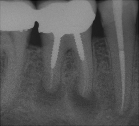
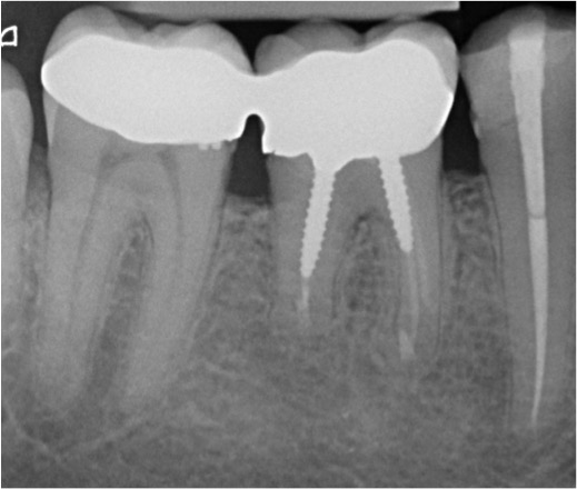
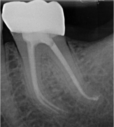
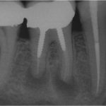
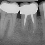
Pre-operative X-Ray where the incomplete sealing of the channels of a lower left molar and the presence of granuloma in the distal root are highlighted.

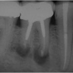
Subsequently the radiography at 1 year after endodontic reprocessing highlights the perfect sealing of the canals and the disappearance of the granuloma.

Discover the details